INTRODUCTION
Mammography
is one of the most demanding radiologic techniques. It requires an excellent spatial resolution
to allow visibility of microcalcifications and good contrast sensitivity to
allow detection of breast tumors. It is
difficult to visualize very small physical changes in breasts with general
x-ray imaging. That is why mammography
equipment was designed differently from general x-ray.
UNIQES FEATURES OF MAMMOGRAM TUBE
Mammography equipment consists of
two major components. They are an x-ray
tube and an image receptor. The x-ray
beam originates at the x-ray tube and transmitted through the breast. A film contained in the image receptor to record
the images from x-ray distribution that passed through the breast tissue.
|
Image
receptor
|
Figure
1. Mammogram
Equipment. Reprinted from Screen-film Mammography Equipment Unit 3, by B.A.
Barnes & X. Ho, n.d., Retrieved from www.santarosa.edu/.../Unit%203%20-%20Mammography. Reprinted with permission.
Features of mammogram x-ray tube
that are unique compare to general x-ray tube include x-ray tube anode, target
materials, filament design, filter, focal spots, source-to-image distance, and
object to image receptor distance.
Most x-ray tubes use tungsten as the
anode material, however mammography equipment uses molybdenum and in some
designs, it uses a dual material anode with an additional rhodium track. Molybdenum and rhodium are used because they
produce a characteristic radiation spectrum that is close to optimum for breast
imaging (Sprawls, 1995).
To reduce unnecessary exposure to
the patient, most x-ray machines use aluminum to filter the x-ray beam. Sprawls (1995) mentioned that mammography
uses filters that work on different principle and also used to enhanced
contrast sensitivity. Same as in the
anode, molybdenum is the standard filter material. Some systems allow the operator to select
either the molybdenum or rhodium filter to optimize the spectrum for specific
breast conditions (Sprawls, 1995).
There is also exit window filtration
(Barnes & Ho, n.d.). Instead of
glass, beryllium is used in dedicated mammography tube housing. Noted that glass act as a filter when dealing
with this soft end of the x-ray spectrum, and it filters out photons that would
not provide contrast (Barnes & Ho, n.d.).
Mammographic x-ray tubes typically
have dual filaments in a focusing cups that produces 0.3 and 0.1 mm nominal
focal spot sizes (Barnes & Ho, n.d.).
Meaning x-ray tube for mammography has two selectable focal spots. The spots are smaller compared to other x-ray
procedures because mammogram requires for minimal blurring and good visibility
of detail to see the small calcifications.
The smaller of the two spots is generally used for the magnification
technique (Sprawls, 1995). Magnification
views use a small magnification table which brings the breast closer to the
x-ray source and further away from film plate.
This allows the acquisition of zoomed in images of the region of
interest (special mammography views,
2008).
Other unique component in
mammography tube which is source-to-image distance (SID). SID is the entire distance of the x-ray beam
from the focal spot to the image receptor (Barnes & Ho, n.d.). The greater the distance the less geometric
blurs occur. This affects the focal spot
size which affects the size of the objects being imaged. The larger the SID the larger the field
size. With larger distance of the source
to the image, more beam will be needed to penetrate which then can increased
the heel effect (Barnes & Ho, n.d.).
Other feature is object to image
receptor distance (Barnes & Ho, n.d.).
It is the distance of the object (the breast) to the image receptor. Increase of the object to image receptor
distance will increase magnification of an area of breast tissue. It will increase the resolution and contrast
of the breast image (Barnes & Ho, n.d.).
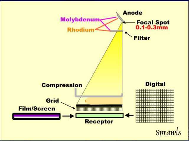
Figure
2. Principle of Mammogram Tube. Reprinted from Mammography Physics and
Technology for Effective Clinical Imaging, by P. Sprawls, 1995, Retrieved from http://www.sprawls.org/resources/MAMMO/mammo02.jpg. Copyright 1995
by Sprawls Educational Foundation. Reprinted with permission.
THE X-RAY SPECTRUM
The x-ray spectrum depends on the combination of three factors. First factor is the x-ray tube anode material
(molybdenum or rhodium), second is material used for x-ray beam filtration (molybdenum
or rhodium), and third is the kV which ranging from 24 kV to 32 kV (Sprawls,
1995).
As for x-ray tube anode, most
mammogram unit uses molybdenum anodes.
However, in some systems have a dual-track anode that allows
radiographer or AEC system to select between molybdenum and rhodium.
For
x-ray beam filtration, the material used is also molybdenum but in some systems
they have an alternative rhodium filter that can be selected. According to Sprawls (1995) the molybdenum
filter only should be used with the molybdenum anode. However, the rhodium filter can be used in
combination with both the molybdenum and rhodium anodes.
Figure 3. Factors
Affecting the X-ray Spectrum. Reprinted from Mammography Physics and Technology
for Effective Clinical Imaging, by P. Sprawls, 1995, Retrieved from http://www.sprawls.org/resources/MAMMO/mammo18.jpg.
Copyright 1995 by Sprawls Educational Foundation. Reprinted with permission.
In physics, there are two types of
x-ray radiation produced when electrons hit the x-ray tube anode. They are bremsstrahlung and
characteristic. Bremsstrahlung is in the
form of a broad continuous photon energy spectrum with a maximum energy
determined by the selected kV value (Sprawls, 1995). Characteristic radiation is produced under
certain conditions and is confined to just a few photon energies (Sprawls,
1995).
The photon energies of the
characteristic radiation are determined by the atomic characteristics of the
anode material which varies with the atomic number (Z) of the material. For mammography, molybdenum and rhodium are
materials that produce characteristic x-ray radiation that is near the optimum
energy which is why they are used for the anodes (Sprawls, 1995).
In mammography, the filters used are
based on the ‘k-edge’ principle and it attenuates the radiation above the
k-edge energy of the specific material (Sprawls, 1995).
Figure 4. K-edge
Principle. Reprinted from Mammography Physics and Technology for Effective
Clinical Imaging, by P. Sprawls, 1995, Retrieved from http://www.sprawls.org/resources/MAMMO/mammo28.jpg.
Copyright 1995 by Sprawls Educational Foundation. Reprinted with permission.
Sprawls (1995) noted that with an
atomic number of 42, molybdenum has a k-shell binding energy and its k-edge at energy
of 20 keV and rhodium, with an atomic number of 45 also has a k-shell binding
energy and its k-edge at energy of 23.22 keV.
When molybdenum filter is selected, it attenuates and blocks much of the
bremsstrahlung spectrum above the energy of 20 keV and gives results in the
spectrum that is most often used in mammography which is ‘moly/moly’
anode/filter combination (Sprawls, 1995).
Figure 5. The
Moly/Moly Spectrum. Reprinted from Mammography Physics and Technology for
Effective Clinical Imaging, by P. Sprawls, 1995, Retrieved from http://www.sprawls.org/resources/MAMMO/molymoly.jpg.
Copyright 1995 by Sprawls Educational Foundation. Reprinted with permission.
The k edge boundary is shifted to a
higher energy with the rhodium filter.
So the portion of the bremsstrahlung between 20 keV and 23.22 keV is
added to the x-ray beam. This makes the
beam more penetrating which provides some advantage when taken image of larger
or denser breast (Sprawls, 1995).
Figure 6. The
Moly/Rhodium Spectrum. Reprinted from Mammography Physics and Technology for
Effective Clinical Imaging, by P. Sprawls, 1995, Retrieved from http://www.sprawls.org/resources/MAMMO/molyrho.jpg.
Copyright 1995 by Sprawls Educational Foundation. Reprinted with permission.
Another anode material is
rhodium. Rhodium can be selected to
produce a more penetrating x-ray beam. Rhodium’s
atomic number (Z) is 45 and has principal characteristic radiation at energy of
20.3 keV with a less intense emission at 22.7 keV. Compare to molybdenum which atomic number (Z)
is 42 and principal characteristic energy of 17.6 keV with less intense peak at
19.7 keV (Sprawls, 1995).
The rhodium filter, with a k-edge
cut off at 23.22 keV, is always used with the rhodium anode. The molybdenum filter cannot be used with
rhodium anode because its k-edge cut off from 20 keV upward would attenuate the
rhodium 20.3 keV and 22.7 keV radiations (Sprawls, 1995).
Figure 7. The
Rhodium/Rhodium Spectrum. Reprinted from Mammography Physics and Technology for
Effective Clinical Imaging, by P. Sprawls, 1995, Retrieved from http://www.sprawls.org/resources/MAMMO/rhorho.jpg.
Copyright 1995 by Sprawls Educational Foundation. Reprinted with permission.
Third factor that affect the x-ray
spectrum is kV. Increasing the kV has
two effects on the x-ray beam. First, it
increases the efficiency and output for a specific mAs value and second it
shifts the photon energy spectrum upward so that the beam becomes more
penetrating (Sprawls, 1995). Penetrating
beam reduce contrast sensitivity and it is necessary for dense breast. Therefore compressed breast thickness is the
principal factor that determines the optimum kV (Sprawls, 1995).
Figure 8. KV
Selection for Different Breast Thickness. Reprinted from Mammography Physics
and Technology for Effective Clinical Imaging, by P. Sprawls, 1995, Retrieved from
http://www.sprawls.org/resources/MAMMO/mammo32.jpg. Copyright 1995 by Sprawls
Educational Foundation. Reprinted with permission.
DISCUSSION
According to Sprawls (1995) the
photon energy spectrum of the x-ray beam is one of the most critical factors in
optimizing a procedure with respect to contrast sensitivity and radiation dose. Although many said that anode materials used
in mammogram can only be molybdenum and rhodium, today many researchers do the
study on tungsten as an anode material for mammogram tube to see whether it
still can produce good contrast and low radiation dose.
Between all of the anode materials
used in mammography tube, which are molybdenum, rhodium, and specialized
tungsten, many researchers agree that molybdenum is the best material to be
used because it allows production of low energy spectrums of radiation and only
need low kVp which is 26 to 30 kVp (Barnes & Ho, n.d.).
However, recently there is study done
using tungsten as an anode material with rhodium in mammography and according
to researchers, if the molybdenum x-ray tube digital mammography demonstrated a
30 percent reduction in dose, the introduction of tungsten x-ray tubes with
digital mammography allows even greater reduction in radiation exposure,
without affecting image quality (Smith, Chen, & Semine, 2005).
Another study done from Dance,
Klang, and Sanborg (2000) to compare performance of mammographic x-ray systems
that use different anode/filter combinations for screen film and digital
imaging. For screen film mammography,
result for thicker breasts shows 20 percent improvement in contrast can be
achieved but without reduce radiation dose using molybdenum/rhodium or
rhodium/rhodium, Whereas more than 50
percent of dose saving can be attained but no improvement in contrast using
tungsten/rhodium or rhodium/aluminum spectra.
As in digital mammography, Dance et al. (2000) mentioned that
molybdenum/molybdenum spectrum delivers the lowest dose for a two centimeter
breast, but gives the highest dose for thicker breasts. However tungsten/rhodium or rhodium/aluminum
spectra provide the lowest doses at greater thickness. Researchers concluded that from this study,
molybdenum/molybdenum is the spectrum of choice for all but not for thicker or
most glandular breasts.
CONCLUSION
The most important part of dedicated
equipment in mammography is the x-ray tube.
The tube is designed and constructed uniquely and specifically for
imaging the soft tissue of the breast.
The x-ray machines used for mammograms today designed to produce lower
energy x-rays but improves in image quality and less radiation.
REFERENCES
Barnes, B.A., (n.d.).
Screen-Film Mammography Equipment Unit 3. Retrieved April 27, 2013, from www.santarosa.edu/
Dance, D.R., Klang,
A.T., Sandborg, M., Skinner, C.L., Smith, A.C., & Carlsson, G.A. (2000). Influence of anode/filter material and tube
potential on contrast, signal-to-noise ratio and average absorbed dose in mammography. The British Journal of Radiology, 73 (2000), 1056-1067. Retrieved from
bjr.birjournals.org/content/73/874/1056.full.pdf
Smith, A., Chen, B.,
& Semine, A. (2005). Minimizing Dose in Digital Mammography. Retrieved May 02, 2013, from
www.hologic.com.data/WP_00005_tungsten
Sprawls, P. (1995).
Mammography Physics and Technology for Effective Clinical Imaging. Retrieved April 28, 2013, from
www.sprawls.org/resources/mammo/module.htm
Special
Mammographic Views. (2008). Retrieved April 28, 2013, from www.imaginis.com/mammography/special-mammography-views-spot-c
No comments:
Post a Comment